Posts

Semester 2 Week 13 - Brinkley

Image
 This week in class the glaucoma gals did the pig head surgery. In this we were able to see what it was like to work on an eye in its actual socket. This added an addition step of positioning the eye to where we wanted it with a suture. This simulation proved to be very educational and will be a cool thing to talk about in an interview.  The pig arrived at the post in a large box with packing peanuts, Alex went earlier in the day to pick it up, so we would have it in time for class.  The pig head being laid out on the tray, getting ready to preform the surgery. Positioning the eye with the suture to allow us to be able to place the valve in the proper spot.  Suturing the valve into place Once the eye was released back into position.  After the surgery we cleaned up the lab and went to the dumpsters to dispose of the pig head waste. The rest of the week was spent solely on the powerpoint. 

Semester 2 - Week 13 Kierstin

Image
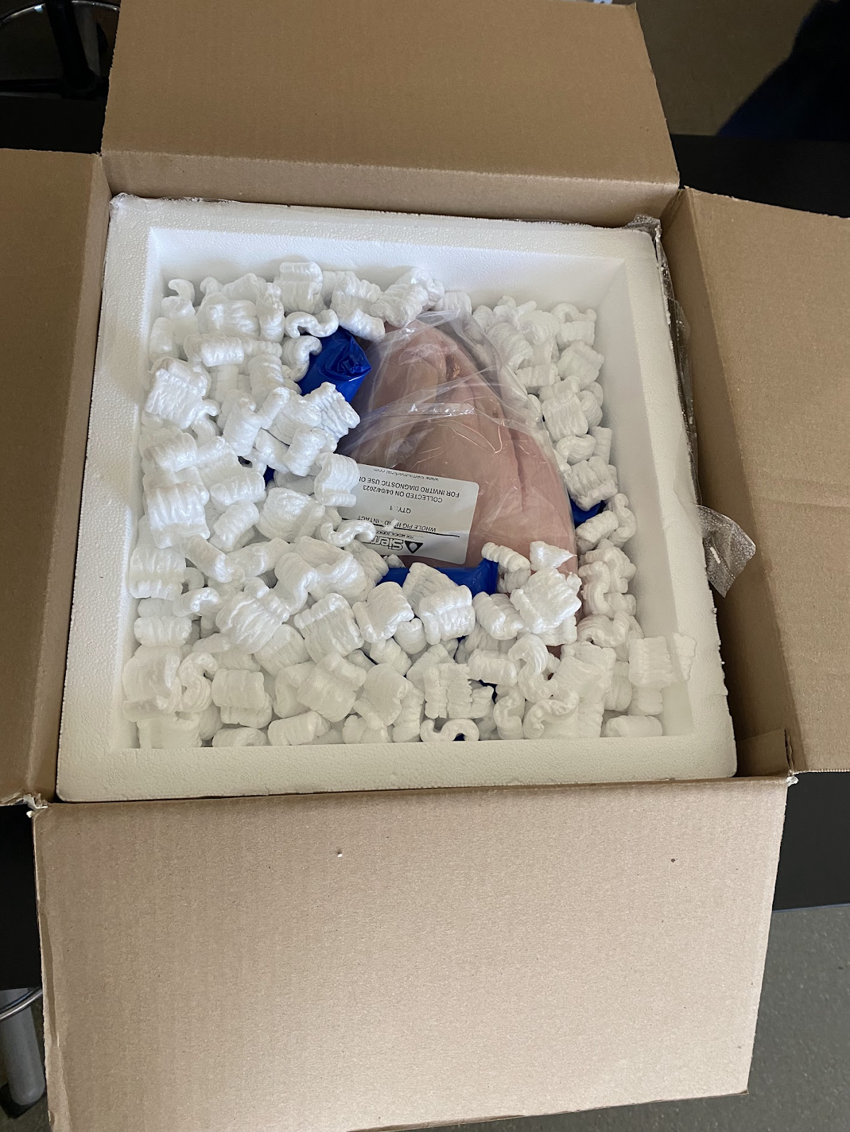
  Lab time:  This week we did our pig head surgery simulation for our verification testing on surgical procedures, and if the device is mobile or not.  Our pig arrived in a box full of packing peanuts. It was very heavy and surprisingly very clean.  We unpackaged the head and placed it in our pre-prepared surgical location. We used trays and covered the table with sticky paper so it would be easy to clean, and nothing would get pig blood on it if it wasn't supposed to.  We started our surgery by suturing the eye so it looked away and giving us a clear spot to cut for the valve insertion. We put the suture through the sclera and then tied it around his ear so it wouldn't move during the surgical procedure.  We then cut the conjunctiva and created a pocket for the valve. I did the main cutting, Brinkley took pictures, and Alex helped give tools and be an extra hand in the surgery. We used scissors to cut it, and then players to create the pocket.  We inserted the valve into the p

Semester 2 Week 12 - Brinkley

Image
 This week in class we discussed with Gordon what needed to be changed and updated on the poster. The notes he had for us were to better explain the reasoning for the three valves and what issues they solve. We updated the poster with these images and explanations.  During lab time, data was collected on our prototype. We were able to find when the 3 valves open and at what pressures. Valve 3 seems to have a defect because even at extremely high pressures it does not open. This is not a fault in our design, but is caused by using faulty Ahmed valves. Here is the data that we collected from class.  Kierstin and I had a meeting on what needed to get changed in the poster, and here are the meeting notes:  After the poster was finished, I began working on our final presentation. I looked over the requirements and began creating an outline for the flow of the presentation. I also worked on finishing my research paper. Because some of my sources were deleted that I researched, I had to go ba

S2W12-Alex

Image
  Data from benchtop testing 

Semester 2- Week 12 Kierstin

Image
  Lab testing:  This week we tested our manufactured valve to see the opening pressures for each ahmed valve in the system. We labeled the valves as follows: 1(left), 2(right), 3(middle)  We used a water column at different heights to test at what pressure mmHg the valve would open and allow flow. The results and lab setup are shown below.  We manufactured another valve that we will be surgically implanting into the pig head when we do that surgery. The valve has the same dimensions as the baerveldt valve, but it is not a functioning valve.  Capstone Poster: A lot of this week was spent working on the poster. I looked over Dr. Gordon's suggestions and made a mock-up of what I was thinking for a timeline of our design. I discussed it with Brinkley to make sure it accurately represented what we thought our product would do over time.  I created these images of the eyes with our specific valve, which took a lot of time because I’m not great at photoshopping things and I had to just us

Semester 2 Week 11 - Brinkley

Image
       Before I went to capstone on Wednesday, Kierstin, Dr. Rickard and I did a demonstration where we inflated and deflated an eye with the pressure from a water column when raised. This helped we gain better understanding of the eye in general and what increased pressure in the eye actually looks like.       In class time we performed multiple eye surgeries. The main goal for this testing was to ge the inlet tube into the eye. The eye was poked with a needle, and the small tubing was inserted, but this proved to be very difficult because of the flexibility in the tube as well as the small size. Dr. Rickard gave us some tips including have the tube cut on a slant to create more of a point for the insertion. He also sad to spin the needle around to create a slightly larger hole than necessary. With his advice we were able to get it. We then disposed of the eyes into the dumpster.  The rest of my time was spent on the poster. In the coming weeks we will have time to make any revisions